1974
Light and electron microscopic studies of the ascus top in Ascozonus woolhopensis
Publication
Publication
Persoonia - Molecular Phylogeny and Evolution of Fungi , Volume 8 - Issue 1 p. 23- 32
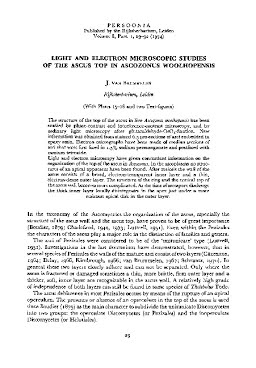
The structure of the top of the ascus in live Ascozonus woolhopensis has been studied by phase-contrast and interference-contrast microscopy, and by ordinary light microscopy after glutaraldehyde-OsO4-fixation. New information was obtained from stained 0.5 μm-sections of asci embedded in epoxy resin. Electron micrographs have been made of median sections of asci that were first fixed in 1.5% sodium permanganate and postfixed with osmium tetroxide. Light and electron microscopy have given concordant information on the organization of the top of the ascus in Ascozonus. In the ascoplasma no structures of an apical apparatus have been found. After meiosis the wall of the ascus consists of a broad, electron-transparent inner layer and a thin, electron-dense outer layer. The structure of the ring and the conical top of the ascus wall becomes more complicated. At the time of ascospore discharge the thick inner layer locally disintegrates in the apex just under a more resistant apical disk in the outer layer.
| Additional Metadata | |
|---|---|
| Persoonia - Molecular Phylogeny and Evolution of Fungi | |
| Released under the CC-BY 4.0 ("Attribution") License | |
| Organisation | Naturalis journals & series |
|
van Brummelen, J. (1974). Light and electron microscopic studies of the ascus top in Ascozonus woolhopensis. Persoonia - Molecular Phylogeny and Evolution of Fungi, 8(1), 23–32. |
|